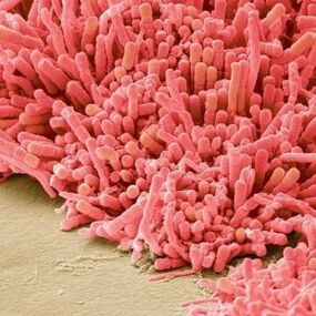
Papilloma is a benign skin neoplasm, the distinctive feature of which is the base of the connective tissue papilla covered with epithelium at the top. Papillomas appear in different parts of the body (skin, mucous membranes, internal organs and other localizations) in humans and most animals.
Papillomas develop from the transitional or squamous epithelium in the form of soft dense formations in the so-called stalk. The size of these formations usually varies from 1 to 2 centimeters, and the outer surface has a white or dirty brown color. Sometimes papillomas grow in different directions and look like cauliflower or a rooster's comb.
Papillomas are removed for cosmetic effects if they appear on visible parts of the body - neck, arms, face, but if they occur in many parts of the mucous membrane, such as the larynx, life-threatening openness can be triggered. In the case of the larynx, papillomas can cause problems with sound by blocking the airways or inability to breathe normally, while in the case of the bladder, papillomas provoke hematuria. If more than one papilloma develops in the body, it indicates the onset of papillomatosis.
Etiology of papillomas
In most cases, the formation of papillomas is caused by a viral infection - human papillomavirus (HPV), although sometimes papillomas can occur as a complication of congenital or inflammatory diseases.
When HPV enters the human body, its activity usually begins to manifest itself after a long time. Often, some stimuli contribute to the activation of papillomavirus, so soft neoplasms begin to appear on the skin or mucous membranes. Among the main factors that provoke papillomas among professionals are stress, decreased immunity, weakness of the body due to treatment, vitamin deficiency in the body, skin damage.
Most people are sexually infected with the papillomavirus, but internal infections are also possible when there are damaged skin areas of the body that have very low immunity or may come in contact with the HPV carrier. The appearance of papillomas indicates the activation of an existing virus, which is equally possible for women and men. A child can be infected with the virus when it passes through the birth canal of an infected mother.

Classification of HPV manifestations
Human papillomavirus infecting mucous membranes and skin can be classified into the following forms:
- Clinical forms that can be detected during routine examination: genital, papular and papillary warts, exophytic warts, as well as cervicitis and cervical erosion in women; Subclinical forms in which the formations are asymptomatic, invisible, and can only be detected by endoscopy: inversions (growing into the mucous membrane), straight warts, as well as condyloma in the cervical canal;
- a latent form characterized by the absence of a clinic and revealed only by the results of tests;
- A female form or cervical form characterized by cervical cancer or dysplasia of various stages.
When women become infected with high levels of oncogenic HPV as a result of sexual intercourse, the likelihood of malignant neoplasms in the cervical canal increases dramatically. When infected with other types of viruses, the chances of oncology are not so high, but a cancerous tumor can form in the rectum or oral cavity. In men, the possibility of cancer due to HPV is present in the anus, penis and rectum.
Types and forms of papillomas
It is very important to correctly identify the papillomas that appear on the body. Their types are directly dependent on the strain of the virus that enters the human body and contributes to the process of excessive cell division in the skin, resulting in the formation of papillomas.
HPV strains can be oncogenic or non-oncogenic. There are more non-oncogenic species and, as a rule, they bring the patient nothing but external aesthetic discomfort.
Such a manifestation can be easily eliminated, and thus solves the problem. However, if neoplasms of the mucous membrane appear, it indicates a serious pathological process. Such an outflow means that a person is infected with an oncogenic HPV strain, so complex antiviral therapy is extremely necessary. To distinguish different types of papillomas, it is enough to compare them with each other or to identify different features of this or that subtype.

Simple warts
Simple papillomas or warts are the most common type of papillomavirus caused by several strains at the same time. These HPV strains are transmitted not only sexually, but also through contact and daily life, leading to statistics showing that 30% of the world's population is exposed to HPV at least once in their lives.
Simple papillomas or vulgar (ordinary) warts are more common in other parts of the upper extremities, ie in the hands, but can sometimes be found on the body, legs and feet, palms and fingers. Their peculiarity is that such warts appear in areas with damaged skin due to reduced local immunity. Such papillomas occur in the lower or palm areas due to contact with poor quality household chemicals, profuse sweating, various skin lesions, dermatitis.
Vulgar warts look like papillary neoplasms of the skin with a diameter of a few millimeters at the beginning of the disease from the outside. In this case, the wart head has a homogeneous and soft texture and rises above the surface of the skin. It is weakly pigmented and penetrates deep into the skin, where the root is nourished by bark. As a result of this diet, warts gradually grow, changing not only their size, but also the degree of pigmentation. Also, hair often grows in the center of such papillomas, which is a variant of the norm and does not show a malignant neoplasm.
Straight papillomas
Similar skin lesions look like small yellowish flat plates that rise slightly above the surface of the skin. The structures are dense, with a deep subcutaneous root, often evidenced by pain when pressing the wart or being damaged in daily life. The localization of such papillomas is most often the face and hands. It can sometimes occur in the anus or in the labia majora in women and in the scrotum in men. There is an active upward trend due to the active blood supply.
The main feature of flat papillomas is the difficulty of their treatment. After surgical treatment of these neoplasms, wounds and scars usually remain in place.
Genital warts
Genital warts occur in the groin area or mucous membranes. Externally, these are thin papillary neoplasms 2-3 millimeters in diameter. Such condylomata grow rapidly, forming a large papillary-like skin growth resembling a cauliflower or a rooster.
The main danger of genital warts is the high risk of infection, inflammation of neoplasms in the vagina or labia in women. They can be easily injured, after which the infection penetrates the body at high speed. In addition, a major problem associated with genital warts is the high risk of recurrence, which does not decrease even with the use of antiviral therapy and removal of neoplasms. Several types of the virus can cause genital warts, and some can be dangerous for women in terms of the malignant process.

Filiform papillomas
Threaded papillomas with a thin stalk crowned by the head of the upper neoplasm. Due to their special appearance, it is very difficult to confuse with other species, so looking at the photo of filamentous papillomas, they may differ from other species.
Such neoplasms appear most often after the age of 45 in areas dominated by thin skin - chest, armpits, neck. The increase in the size of such neoplasms is their further elongation. The head of filamentous papillomas is usually yellowish or pink, pigmentation is not expressed, in most cases very weak.
Domestic goods
Any neoplasm on the surface of human internal organs can be classified as a subgroup of internal moles. These are intracranial condylomata, papillomas in the rectum, neoplasms in the throat and mouth, and neoplasms in the walls of the bladder. A distinctive feature of these papillomas is the impossibility of their recognition without appropriate medical procedures and diagnoses. However, the disease may be suspected with specific symptoms. The danger of such neoplasms is determined in each case.
If there are papillomas in the bladder, bleeding or cancer may develop over time.
If the papilloma is in the larynx, it prevents breathing and interferes with a person's speech function.
Lewandowski-Lutz papillomas
Warty epidermodysplasia, or Lewandowski-Lutz papilloma, is a very rare pathology that mainly affects only children or adolescents. Such a disease can be inherited and spread in a family.
The clinical picture of the disease manifests itself in the form of numerous reddish-brown spotted warts on the feet and hands. One of the features of the pathology is that when papillomas are located in areas of the body exposed to ultraviolet radiation, in one third of all cases they again turn into malignant neoplasms and the growth of neighboring tissues.

localization of papillomas
Filamentous, vulgar or pointed papillomas, as well as condylomata are the most common in medical practice. Filamental warts are localized, vulgar ones are more common in the legs or hands, and condylomata are found only in the mucous membranes (head of the penis and urethra in men, small labia and vagina in women), but these warts may be unusual. can.
It is not difficult to remove such papillomas in modern conditions, but the danger is that the re-emergence of new papillomas with reduced immunity will lead to more serious health consequences, for example, the subsequent development of genital warts in women is fraught with cervical cancer. childhood. Plantar warts are most commonly seen on rough feet and toes. Sometimes a thorn can form on the thumb after severe damage to the skin in the area.
In general, papillomatosis is a generalized pathological form in which neoplasms form throughout the human body. These growths have a characteristic appearance, so once you see the manifestations of the disease, it can no longer be confused with another disease.
HPV symptoms
The most common symptom of papillomavirus in the human body is the formation of papillomas on the skin.
The rest of the symptomatology depends directly on the location and type of disease. Depending on the above symptoms, the symptoms of HPV can be as follows:
- Genital warts occur on the mucous membranes of the genitals, mouth, throat, rectum and inner surface of the stomach. Symptoms of the onset of pathology in the genital area are itching and an unpleasant odor. If such symptoms become disturbing, they should never be ignored, as they can often be oncogenic.
- Intraductal papillomas in the ducts of the mammary glands with symptoms of redness, slight itching and burning in the nipple. Also, if you press on the tip of the nipple with such a papilloma, an ichor or green discharge begins to leak from it. The danger of intraductal papilloma is a gradual and possible degeneration towards breast cancer.
- Plantar warts are expressed in active calluses in the heel area that cause severe pain when walking or pressing on it.
- Papillomas in the larynx are not initially expressed in any specific symptomatology, but gradually lead to a change in the voice of a person with this pathology, a feeling of coma in the throat and impaired respiratory function. At the same time, the patient begins to have difficulty swallowing.
- In adolescents, flat warts are most common on the outside of the hands and lower face. The symptomatology is very vague and is expressed in mild, rare itching of most neoplasms.

Pathogenesis
When HPV is present in the human body, it is possible to conclude that the immune system is weakened. Once in the body, the viruses begin to infect the basal epithelial layer and tend to affect the area of transition from the squamous epithelium to the cylindrical. Infected cells can contain 2 forms of the virus - benign in nature with episomal (outside the cell chromosomes) and malignant in nature with intrasomal (integrated into the cell genomes).
The incubation period of the papillomavirus can vary from 14 days to one to two years from the time the virus enters the body until the first manifestations of the disease. The nature of human papillomavirus infection is generally secretive. At the same time, several types of pathology can be located in the human body at the same time, and under the influence of certain factors, each of them can begin to manifest itself at the same time through active reproduction. In this case, a stage emerges in which the clinical manifestations of the disease begin to be identified.Very often (up to 90% of all HPV infections) the human body heals itself from this pathology within 6-12 months, but in the remaining 10% of cases the disease becomes chronic with a long course, recurrence and the possibility of malignancy of the processcan.
Diagnosis of the disease
Ultrasound for papillomas
When diagnosing papillomas, ultrasound is not used as the main research method, but as an additional method to confirm the accuracy of the alleged diagnosis. Ultrasound is mainly used to identify papillomas when it comes to malignant transformation in the internal organs.
Ultrasound is one of the instrumental methods used to diagnose intraductal papilloma.
In this case, ultrasound examination does not allow the specialist to examine the mammary ducts, but helps to distinguish intraductal papilloma from suspected breast cancer, excluding galactorrhea in prolactinoma. In addition, ultrasound can contribute to the appearance of neoplasms with bladder papilloma. However, in this case, ultrasound is effective only when the diameter of the neoplasm is more than 1 cm.

PCR diagnostics at diagnosis
The disease is diagnosed by doctors, dermatologists and venereologists. Because the number of virus types varies, it is important to determine exactly what type of virus the patient is infected with and whether the strain is oncogenic. Visually, it is possible to make an accurate diagnosis only in classic genital warts, so if HPV infection is suspected, specialists always use PCR forty.
Polymerase chain reaction (PCR) researchers are not only invited to determine the presence of HPV in the body, but also demonstrate the type, oncogenicity and number of viruses at the time of diagnosis. This is very important from a diagnostic point of view, because if there is information about the percentage of the virus in the body, it is possible to determine the approximate history of infection and identify the patient's contacts for etiotropic therapy.
Based on the results of PCR diagnostics, it is possible to determine the chronic course of the infection or a single flare-up due to decreased immunity. This information allows the specialist to prescribe therapy appropriate to a particular case. Generally, PCR diagnosis is performed in the form of screening. If the presence of a virus in the body is confirmed, the patient continues to be examined using other methods.
HPV biopsy
In medicine, a biopsy is a procedure of taking human tissue samples for further examination by staining them with special dyes. Biopsy is also very common for cancer, such as suspected HPV. Prior to treatment for papillomavirus, physicians should rule out the oncological nature of neoplasms.
Biopsy is a very accurate diagnostic method that can be expressed in cytological or histological studies if HPV is suspected.
A cytological study is a study of body cells under a microscope designed to demonstrate to experts the changes caused by a viral infection in these cells. Cells are taken from this organ for cytological examination in a woman for the prevention and early detection of cervical cancer. If oncogenic HPV is detected in such women in the absence of external manifestations and symptoms, cytological examinations are scheduled each year to allow them to see the signs of cervical dysplasia in a timely manner. The fact is that the dysplasia of this organ is completely cured, and if you do not begin to develop the process, then cervical cancer in the body will not develop, even with an oncogenic type virus.
To make an accurate diagnosis of HPV, a histological examination of the patient is performed, not a superficial cell shear for analysis, but a piece of tissue taken to allow the correct placement of cell layers, tissue characteristics, and oncological features. The tissue sample taken during histological examination with the help of solutions is dehydrated and placed in paraffin, after which the sections are used to obtain layers with a thickness of 0. 1 mm using a microtome. The removed layers are stained with special dyes to detect pathological cells and determine their nature during microscopic examination.

Treatment of papillomatosis
Papillomavirus is always treated on an individual basis. If the virus is detected at the time of diagnosis, but there is no manifestation yet, the patient is prescribed etiotropic cytostatic therapy, which effectively "lulls" the virus for several years.
If a person is an HPV carrier, he or she should have a regular PCR diagnosis to determine the early signs of the disease. In addition, the carrier of the virus is obliged to use barrier contraception to prevent sexual partners.
When papillomavirus is detected, the use of antiviral drugs in treatment is mandatory. In general, immunomodulators and vitamin supplements are indicated for all patients with HPV.
Depending on the location and symptoms, when papillomas form on the mucous membrane or on the skin, cryodestruction, electrocoagulation and laser removal of the growths are used. Sometimes papillomas are removed with a more modern technique - using radio waves. When there are signs of papilloma malignancy, it is surgically removed along with the healthy tissue around the growth. It is also important to know that the removal of a papilloma does not lead to a complete cure, because the virus remains in the body and can recur.
Because papillomavirus is the most sexually transmitted virus, a barrier method of contraception should be preferred, and if a woman is planning a pregnancy, it is important to take timely diagnostic measures and provide therapy that will reduce the chances of the child becoming infected with the virus.In modern medicine, there is no drug to completely eliminate the virus from the body, so when such a diagnosis is made, even in the absence of manifestations, a person should be regularly examined to detect the development of pathology.
Disease prevention
It is possible to prevent papillomas in the body by following the basic rules of hygiene and timely disinfection of wounds. In daily life, it is important for each family member to use a separate towel, a comb, manicure tools, shoes, and inappropriate sexual intercourse should always be protected with a condom. It is also important to take regular showers after sex and treat the contact areas of the skin and mucous membranes, as it takes some time for the virus to enter the human body.
In modern medicine, there is a vaccine against human papillomavirus. It has already been tested in 72 countries around the world, and is effective against HPV subtypes 16 and 18, which cause cervical cancer in 90% of all diagnosed cases. In addition, vaccination successfully fights subtype 6 and 11 viruses that cause the development of genital warts, which are difficult to treat. Due to the sexual transmission of these viruses, it is recommended that a person be vaccinated before sexual activity. Experts often recommend that the vaccine be given three times a day to girls between the ages of 11 and 12. The World Health Organization recommends that boys be vaccinated to prevent the possibility of HPV.
Are papillomas dangerous?
Papillomavirus is a risk factor for the development of oncological pathologies. The virus often causes cervical cancer, cancer of the external genitalia (vulva, glans penis). However, HPV infection does not always lead to cancer. There are many subtypes of this virus with a low oncogenic index, such as subtypes 6, 11, 42, 43, 44, which cause condyloma, as well as highly oncogenic subtypes that cause flat warts - 16, 18, 31, 33. The virus enters the bodyIt can take 10 to 20 years from the time it enters to the transformation of a neoplasm into a malignant disease.
If there are large papillomas in the body that can be easily damaged in daily life, they should be removed.
If left untreated, the risk of contracting other infections increases dramatically. With the passage of parallel infectious processes, papillomas begin to appear in other parts of the body and weaken the immune system. The massacre turned out to be a circle. In addition, if some papillomas are not removed, they can turn into oncological neoplasms, which means that the disease should be treated with all seriousness and never allow the disease to progress.


